Safety Assessment
|
Regina Kelder
A “Liver-on-Chip” for Genotoxicity Evaluation?
Scientists from Charles River are gaining ground in finding an alternative to the rodent model using rapidly advancing 3D-culture technology
Thalidomide is a well-known cautionary tale about why it is important to test drugs for safety. The sedative developed in the 1950s as a treatment for morning sickness in pregnant women was withdrawn in 1961 after it was finally established that the medicine was responsible for babies being born with underdeveloped arms, legs, and other malformations.
This tragic chapter led to the worldwide adoption of more rigorous testing procedures and regulations stipulating that drugs should be tested for potential side effects on the fetus, using mice, rats, or rabbits.
Today, drugs and chemicals cannot move forward without a slew of animal safety tests including genotoxicity evaluation, which are designed to detect drugs and other substances that have the potential to alter or damage our genetic material and lead to cancer. Genotoxicity can lead to indirect or direct effects on the DNA including inducing mutations that can be passed to future generations of cells.
Genetic toxicity tests are not just important for pharmaceuticals. They are also used on chemicals, medical devices, cosmetics, and textiles. For decades laboratories used a battery of tests, either in rodents or in vitro, to measure how toxic substances were to our DNA. Three of the most common genotoxicity assays today are: Comet assay, which measures DNA strand breaks in cells; the Ames test, which uses multiple bacterial strains to detect genetic mutations following exposure to a chemical or other test material agent; and micronucleus test, a well-established assay that quantifies DNA damage. Many genotoxicity studies use multiple methods in conjunction with each other to build up a complete picture of the genotoxic potential of a chemical substance.
While the tests help to prevent another “thalidomide incident” by protecting people from being exposed to carcinogenic agents, they also impose a burden on the pharmaceutical industry by erroneously stalling some products identified as genotoxic that are actually safe. The findings from standard in vivo genotoxicity testing methods can also, at times, be difficult to interpret because they have limited capabilities in detecting genetic damage in multiple target tissues. So, scientists continually search for alternative tools and technologies that might be more reflective of the human condition.
Successfully Navigate Genetic Toxicology Testing Requirements
In this webinar, our experts share thoughts on testing strategies and approaches to address adverse genetic toxicology results. Watch to broaden your understanding of genetic toxicology testing so you can bring your drug to market safely, swiftly, and effectively.
Watch the Webinar
A 3D system for genotoxicology evaluation
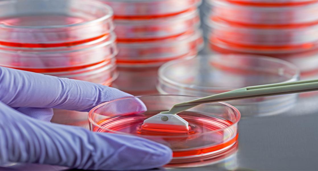
Enter organ-on-chips, a new generation of models that have the capability of complementing and one day replacing rodent models. Organ-on-chips, shown on left, are heavily engineered man-made systems in which the biological components (cells) are subjected to a tissue-specific microarchitecture (engineered part). The tissues are grown inside miniaturized fluid channels molded into glass, silicon, or polymer. The hair-fine microchannels guide and manipulate minute volumes of solution to create the environments that recapitulate one or more tissue-specific functions. Though simpler than human organs or tissues, they are effective mimics of human physiology and disease, which makes them an attractive alternative to the translational challenges of animals.
“All preclinical tests are using mainly animals or rodent-based assays,” says Annie Hamel, Scientific Director of Genetic Toxicology at Charles River’s Safety Assessment site in Montreal. “But all the drugs are going to humans. That is why we are thinking maybe we would have a better predictivity if we were using a humanized model.”
Organ-on-chips are not the only microphysiological system (MPS) being explored as an alternative to convention genotoxicity tests. Spheroids, which are cell aggregates that can form into sphere-like shapes and mimic tissues, are another type of 3D cell culture being evaluated in safety studies. Rounding this out are organoids, which are derived from stem cells (human, and both fetal or adult) that self-organize and originate organ-specific cell types that mimic the structure, function, and cellular complexity of human organs.
Because the liver is the most active organ in metabolism of substances, and frequently a target organ for carcinogens, it makes sense to use a liver MPS to target genotoxicity. But which system?
Hamel said they looked at spheroids as well as a closed liver chip developed by one of the leaders in the development of liver-on-chip technology.
But while closed liver-on-chip systems offer considerable versatility, they are difficult to work with, especially for genotoxicity assessment using life cells, and are also an expensive option so Hamel’s team decided to apply what they learned from the closed chip and apply it to an opened plate well format, which is a more friendly user format, developed by organ chip company CN Bio. The system offered plenty of cells, permeable transwells that can be used to add other types of cells, such as lymphoblasts, and the potential of meeting multiple endpoints required for genotoxicity tests, which currently required multiple studies to be performed.
On the bottom of each well they built a liver organoid that was similar to the closed liver chip, starting with an extra cellular matrix to help the cells attach to the plate, the endothelial cells and then the hepatocytes and a hydrogel on top, so the cells behave as if they were in a real organ.
This model fits all their needs, noted Hamel. “It takes about 7 days to culture the model, which is less than for spheroids, and the media changes are only required every 2-3 days, which is reasonable in term of lab work, and not too time consuming,” says Hamel. “And since it is an open system, it is easy to access the cells, desegregate and prepare healthy single cell suspension just like you would get with healthy tissue.”
An in vitro replacement for rodent tests
Hamel says their main goal is to replace rodent tests with the chip model. “But we will see if regulators accept it and be in agreement with it,” says Hamel. “Currently, cosmetics cannot be tested on animals [in Europe and Canada] so we plan to use this model on cosmetics and maybe chemicals as a start to prove that our system is appropriate, and maybe even better than the in vivo rodent studies. If so, maybe regulators will allow chips to be used for pharmaceuticals, too.”
A recently published proof-of-principle study that Hamel held an overall supervisory role in executing the research at Charles River offers a glimpse of the potential of liver chip technology in identifying genotoxic hazards. It flagged two known carcinogens methyl methanesulfonate and ethyl methanesulfonate that directly damage DNA, and two carcinogens—benzo[a]pyrene (B[a]P) and cyclophosphamide—that require metabolic activation. Findings for the study, which also included researchers from CN Bio and TwinStrand Biosciences in Seattle, appeared in the May / June issue of Mutation Research: Genetic Toxicology and Environmental Mutagenesis.
Hamel said they were currently testing the LC-12 plates specifically designed for liver cells and are currently looking at evaluating the micronucleus test using human hepatocyte cells, rather than the lymphoblast (TK6) cells. “In order to prove it's really a good model that can replace animal testing, we will need to test it in multiple compounds,” says Hamel, adding that typically new assays need to be tested on generally at least 60 different types of compounds. “We will also need to transfer the method to other labs to show that the model is appropriate and build confidence in the results.”
Hamel has directed over 350 in vitro and in vivo safety assessment studies at the Montreal site and has extensive experience working with co-cultured 3D models, so she knows the degree of difficulty in introducing a new assay to a lab. Her lab has been chipping away (pardon the pun) on this project for already more than five years. They are not the only lab looking into organ-on-chip technology, but they are further ahead than most labs in testing liver-on-chips as an alternative to the rodent model.
“It’s time to rewrite the script on scientific progress,” says Hamel. “By embracing the 3Rs of replacement, reduction, and refinement – we can unlock a world of innovation without compromising ethical boundaries. Let’s harness the power of technology and human ingenuity a future where animals are no longer required.