Safety Assessment
|
Russell Garland
The Role of the Immunology CRO Lab
Identifying Infusion-related reactions to monoclonal antibodies in early phase clinical studies can help guide administration and pre-medication strategies
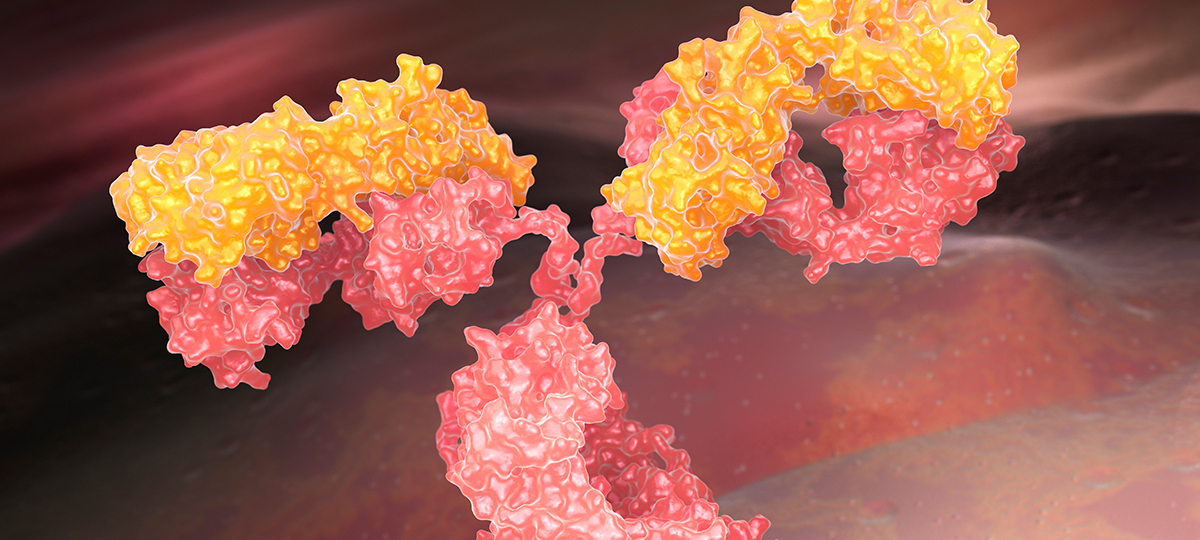
Monoclonal antibodies (mAb) and immune-enhancing agents have become important weapons in the oncologists’ arsenal for the treatment of cancer. Conventional human antibodies as well as bispecifics and modified antibody-based platforms are being used extensively to block inhibitory pathways in the immune system, drive costimulatory signals or directly target immune responses to the tumor cell. Similarly, many programs are now aimed at the generation of small molecule modulators of the same pathways.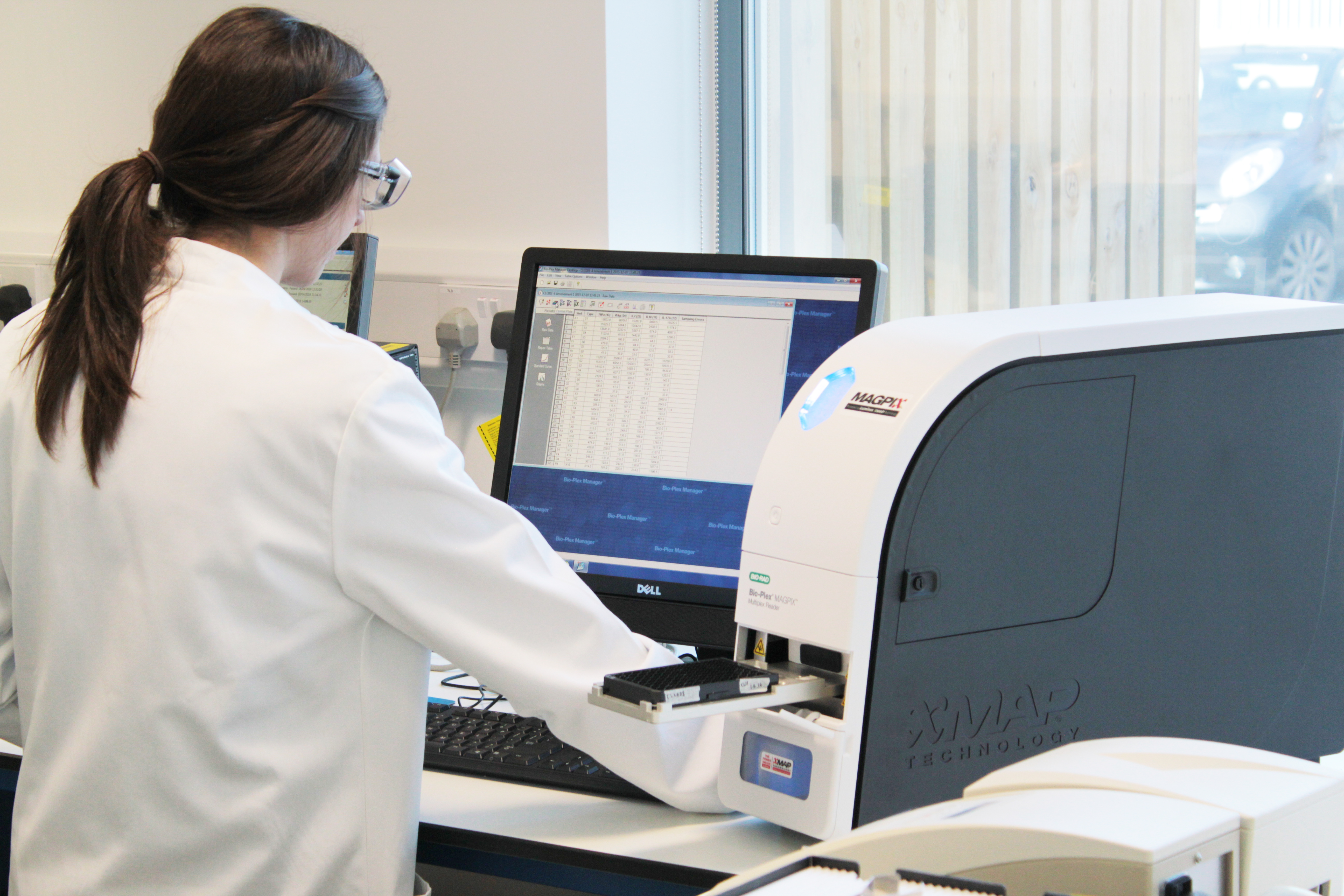
A consequence of enhancing immune reactivity is however the potential to drive unwanted drug reactions. If reactions occur within 24 hours of mAb administration they are considered to be infusion-related reactions (IRRs) and, in oncology patients, are graded according to the Common Terminology Criteria for Adverse Events (CTCAE). Safety monitoring and post-marketing pharmacovigilance generates estimates of the rates and severity of reactions observed to each therapeutic agent. In the clinic, once an IRR is triggered it can progress rapidly, so close monitoring of potential symptoms combined with the administration of an appropriate pre-medication regime (frequently steroids and antihistamines) is important. The underlying immunopathology can involve several immunological mechanisms, which cannot accurately be distinguished by based on clinical symptoms.
Cytokine release syndrome (CRS, acute infusion reaction) is the most common IRR type after mAb administration. Often, CRS IRRs occur 30-90 minutes into the first mAb dose. In the case of mild-to-moderate IRRs, treatment may be tolerated without discontinuation or (after symptom resolution) using a slower infusion rate. For more severe (grade 3-4) IRRs re-exposure is discouraged. CRS have been reported with Rituximab (anti-CD20), which is frequently used in the treatment of leukemias and lymphomas, and after treatment with the CD3/CD19 bispecific antigen binder Blinatumomab and after chimeric antigen receptor (CAR) modified T lymphocytes (although the latter is obviously not a mAb therapy).
Type I Hypersensitivity (allergic reaction) can occur within minutes of a mAb infusion, suggesting some prior sensitization leading to pre-existing IgE which cross-reacts with the mAb upon infusion, or the generation of anti-mAb IgE upon earlier exposure. Unlike CRS, where the severity typically subsides with each subsequent dose, re-challenge is contraindicated for suspected (IgE-mediated) anaphylaxis.
Complement activation-related pseudoallergy (CARPA) is reported as a hypersensitivity reaction which is a consequence of complement system activation (C3a, C5a and C5b-9), rather than IgE antibodies. It typically occurs within minutes after starting the infusion. Most often this occurs on first treatment without prior exposure. Slower infusion rates are suggested for subsequent administrations, and the reactions often subside following repeated exposure.
The reporting of IRR is somewhat confused by the inconsistent use of terms. Given the speed with which these reactions can develop and progress, the laboratory cannot typically provide results indicating the underlying mechanism in time to influence patient management decisions. However, analysis of cytokine, complement and IgE levels in early phase clinical trials can provide important information on the immunopathology type, which can then be used to guide administration and pre-medication strategies. Such analyses also have the important role of identifying cases of IgE-mediated IRR where repeat exposure should be avoided.
Get Support for your immunology research
NOTE: This blog post first appeared on Aug. 22, 2017 in the KWS Biotest blog.