
Lung Infection Models for Preclinical Antimicrobial Drug Development
Bacterial infections in the lung leading to pneumonia are a major cause of death in the young, elderly, immunocompromised and cystic fibrosis patients. The development of multi-drug resistant strains makes effective treatment difficult. Potential novel treatments may be assessed in several models of murine lung infection including Pseudomonas aeruginosa, Streptococcus pneumoniae, Staphylococcus aureus. The duration and severity is dependent on the bacterial strain. Efficacy of client treatments can be determined by reduction in clinical disease, improved survival, and bacterial load in the lung. The lung can also be assessed for gross pathology, cellular infiltrate and cytokines in the bronchial lavage fluid (BAL) and histopathology. For models that replicate persistent infections, see our chronic Pseudomonas lung infection poster.
WEBINAR: Secure Regulatory Approval of Complex Lung Models Through Collective Momentum
Regulatory agencies are increasingly embracing in vitro lung models. By integrating these advanced models, you can generate more human-relevant data, helping you streamline regulatory approvals and improve decision-making.
Watch the Replay
Lung Infection Model Study Endpoints
- Clinical Scores
- Change in bodyweight and temperature
- Bacterial load:
- In life analysis via bioluminescent imaging (IVIS)
- CFU in lung homogenates
- Bioanalysis – cellular infiltrate and cytokines in BAL or lung homogenate
- Gross pathology scores
- Histopathology (H&E staining, IHC)
Tell Us How We Can Support Your Program
Validation Data of Lung Models
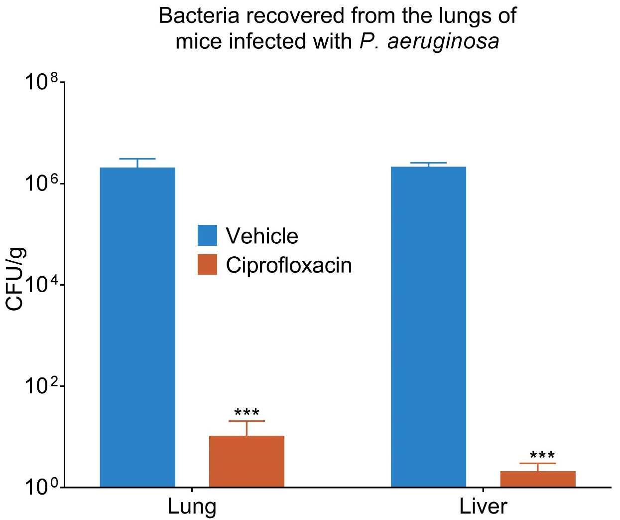
Figure 1. Bacterial load in the lung and liver of untreated and ciprofloxacin treated animals show a significant reduction following treatment.
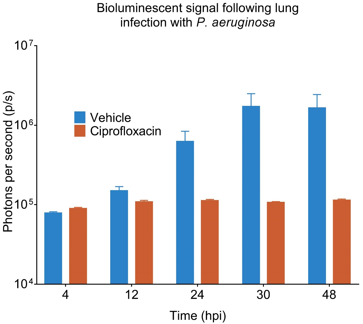
Figure 2. Growth of bioluminescent bacteria in-life can be monitored using IVIS technology.
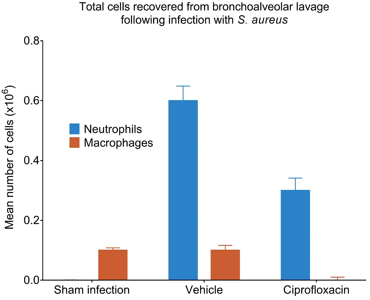
Figure 3. Cellular infiltrate in bronchial lavage fluid can be assessed by FACS.
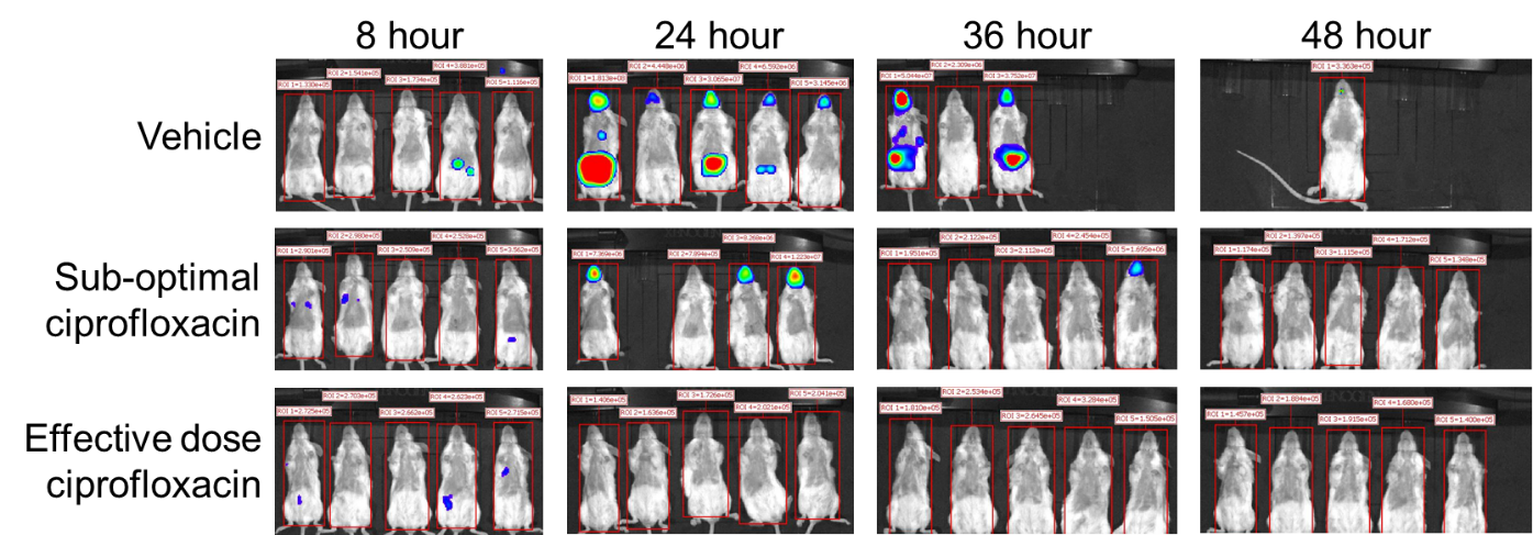
Figure 4. IVIS images at different time points following infection show level of infection.