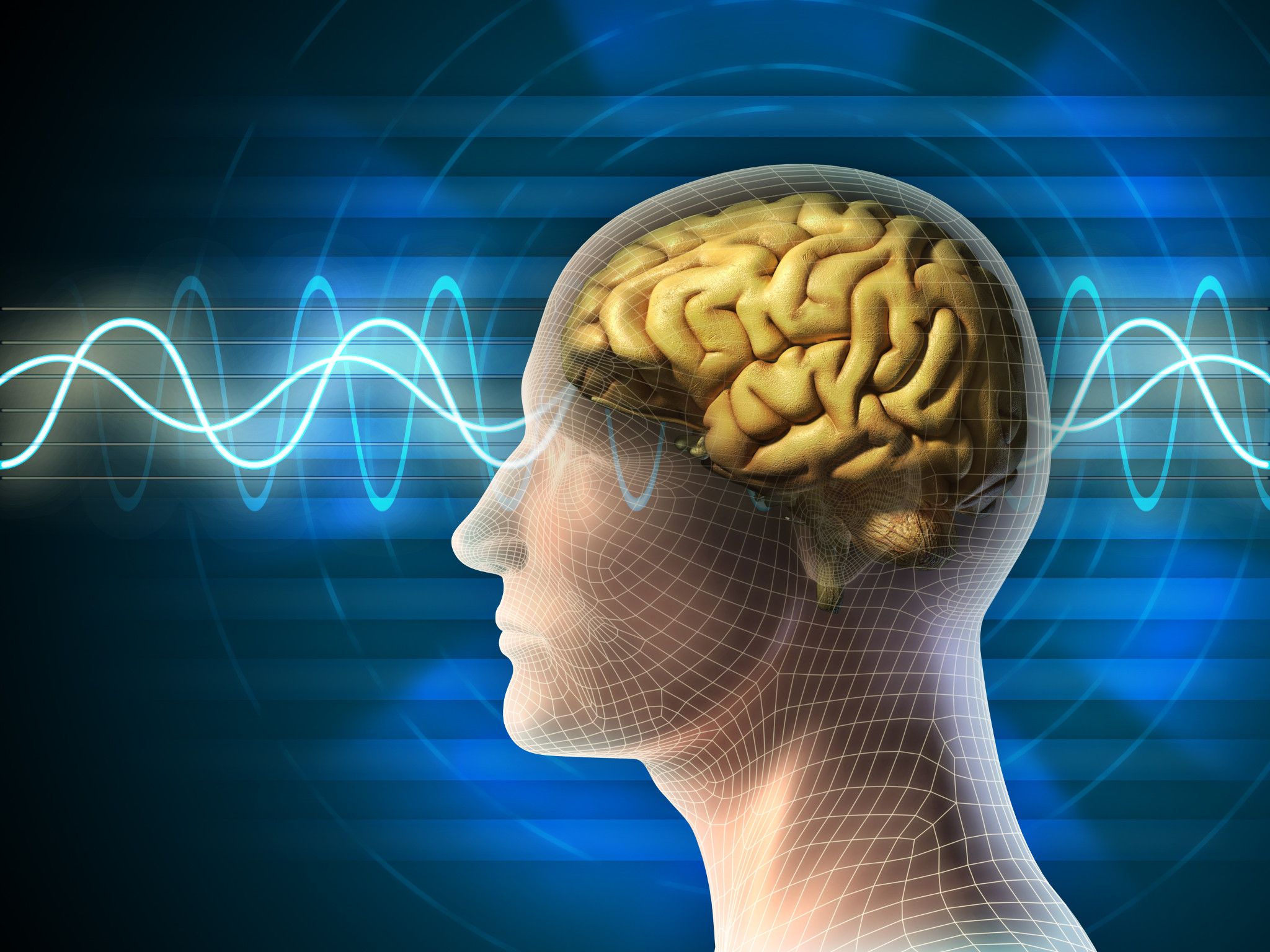
Mouse EEG and EMG Studies
Understanding the effect of novel therapeutics on brain activity and function is vital to demonstrating efficacy in drug development for epilepsy and seizure disorders, as well as assessing the impact on sleep-wake cycles in sleep disorders or other neurological diseases where sleep is altered. Employing mouse EEG and EMG, together or independently, provides quantitative and translatable data to drive your neuroscience drug discovery forward.
See how MEA electrophysiology can be used with primary cells, human iPSC-derived cells, and ex vivo brain slices across different stages of drug discovery in our on-demand webinar.
Watch Now
EEG and EMG drive informed decision-making in drug discovery
Validating the effectiveness of a new therapeutic in your target disease is crucial to drug discovery. Our expert team of scientists can help you interpret complex mouse EEG and EMG data, enabling you to assess the impact of your test article on neuronal and neuromuscular function in relevant disease models.
Electroencephalography (EEG)
EEG is the gold standard for investigating seizures and epilepsy-related tests, providing reliable and objective measurements of electrical activity. Mouse EEG measures the postsynaptic potential of neurons in the outer layers of the cortex as the summed activity of multiple neurons. This non-invasive measurement is a highly translatable method applicable to humans, clinical settings, clinical trials, and preclinical animal models. Rodent EEG data can be analyzed by comparison of distinct EEG frequency bands (e.g., delta, theta, etc.) or directly using a comparative power spectral density approach, generating deeper insight into the comprehensive data.
Electromyography (EMG)
EMG measures the magnitude and timing of muscle activation and is routinely used in nerve conduction studies to detect abnormalities in neuromuscular diseases like amyotrophic lateral sclerosis (ALS), multiple sclerosis (MS), and in nerve injury and neuropathy. The amplitude of measured responses corresponds to the force of muscle contraction. EMG measurements are highly flexible and can be used to compare the activation of specific muscles across individuals, groups, and time points. Similar to EEG, EMG is a highly translatable method, providing consistent readouts across drug discovery models and human clinical settings.
EEG and EMG Services and Readouts
Our neuroscience experts can help you select the most appropriate readouts and models to answer all of your drug discovery inquiries. Explore below to learn about different readouts using EEG and EMG with data examples.
-
Seizure Detection With EEG
Seizures characterize epilepsy and related rare disorders and are caused by abnormal synchronized neuronal activity. Understanding seizure activity and the effect of anti-seizure medications on that activity is key to understanding brain function and profiling novel therapeutics. Leverage telemetry recordings to detect and monitor acute or chronic seizure activity, in combination with video tracking and Racine scoring for seizure characterization.
-
Compound muscle action potential (CMAP) and nerve conduction velocity (NCV) by EMG
CMAP measures the electrical response of muscles to motor nerve stimulation and is the summation of almost simultaneous action potentials from several muscle fibers in the same area. Readouts include the latency of muscle response, response amplitude. CMAP is performed in conjunction with NCV measurements to differentiate deficits or drug effects on nerves versus muscles.
Data below shows the maximal peak-to-peak amplitude of responses recorded following sciatic nerve stimulation in SOD1 mouse model of ALS, showing decreased response with age.

-
Sensory nerve action potential (SNAP) by EMG
SNAP provides information on the activity of sensory neurons and is commonly used in neuropathy models, such as chemotherapy-induced neuropathy/pain and nerve injury models.
-
Sleep/Wake Cycle Monitoring (EEG and EMG)
Monitoring the sleep/wake cycle enables the detection of simulant or sedative effects of novel compounds. Additionally, sleep is known to be disrupted in many neurological and neurodegenerative diseases. For example, in depression, shortened latency to REM sleep and increased REM sleep have been identified. Some anti-depressants are known to suppress REM sleep and cause changes in the sleep/wake cycle that EEG can detect. With our telemetry and data analysis approach, we can continuously monitor sleep activity and the effects of novel therapeutics on sleep patterns using detection and comparison of time spent in different stages:
- Wakefulness
- Slow wave sleep (SWS)/Non-rapid eye movement (NREM) sleep
- Paradoxical sleep (PS)/Rapid eye movement (REM) sleep
- Latency to different stages
- Ratio of REM to NREM sleep
The data below was recorded over a 24-hour period, partitioned into awake and sleep periods. In the awake period, blue indicates quiet wakefulness (29.1% of recording time), while green indicates active wakefulness (20.4%). In the sleep period, blue indicates NREM sleep (45.8%), while green indicates REM sleep (4.7%).

Benefits of EEG and EMG in Drug Discovery
![]()
Translational
EEG and EMG are highly translatable methods used in clinical trial and human health
![]()
Versatile
EEG and EMG data enables versatile readouts, with studies designed to answer your specific questions
Learn how we can support end-to-end epilepsy drug discovery from high-throughput screening to in vivo efficacy and safety.
Watch On Demand
Frequently Asked Questions (FAQs) About Quantitative EEG and EMG Services
-
What system does Charles River use for EEG and EMG telemetry?
Our scientists use the DSI small animal telemetry recording system of cages and transmitters, enabling wireless recording of data in freely moving animals.
-
What are the classic spectral frequency bands identified in EEG data?
The frequency of electrical activity recorded by EEG can be defined as bands that correlate with different states or behaviors:
- Delta - <4Hz. High amplitude waves that indicate slow-wave (NREM) sleep in adults
- Theta – 4 to 7Hz. Drowsiness, idling; commonly identified in brain regions that are not related to the task in human task-based EEG
- Alpha – 8 to 12 Hz. Relaxed wakefulness, predominantly identified in the parietal and occipital areas
- Beta – 13 to 30Hz. Low amplitude waves that are indicative of active thinking and focus
-
How is quantitative EEG different than EMG?
Quantitative EEG monitors the activity of the whole brain. EMG monitors the activity of skeletal muscle fibers. They can be used independently or together to monitor brain activity, neuromuscular activity, and sleep/wake cycles.