
Complex cell models for Neuroscience Drug Discovery
Cell culture models are used in drug discovery in screening studies, lead optimization, in vitro efficacy assays, and in vitro toxicology assays. Traditionally, these cultures employ a single cell type (mono-culture), for example of neurons, microglia, or astrocytes, and can be either isolated primary cells from animal models or human post-mortem tissues, or can be derived from stem cells. However, mono-culture does not accurately represent the complexities of the human brain, nor does it allow for investigation of cell-cell communication that is often a therapeutic target.
To enable a deeper understanding of the interplay between cell types found in the brain, and therapeutic targeting of those connections, we have developed innovative, complex cell culture models, with two, three, or four cell types cultured together. For example, we have established and validated co-culture models of neurons and oligodendrocytes to investigate myelination and multiple sclerosis, and also a neuroinflammatory model using neurons, microglia, and astrocytes.
These models can be combined with various readouts and technologies to better mimic your disease state of interest and improve translational success in drug discovery. Example readouts could include:
- High-content imaging using immunostaining and proprietary algorithms to examine cell phenotype and morphology, quantify marker proteins, and determine co-localization of proteins
- Detection of translational biomarkers such as cytokines or neurofilament light chain (NfL)
- Calcium imaging as a readout of neuronal function
- Treatment with disease relevant or inflammatory triggers, such as LPS or amyloid-β monomers
In this webinar, learn why we need better models of brain processes for neuroscience drug discovery, and how our experts have built complex cell models for myelination, neuroinflammation and neurotoxicity.
Listen Now
Myelination Cell Culture Model
Oligodendrocytes are responsible for myelination of neuron axons to support rapid nerve impulse conduction. Dysfunction of these cells, disrupted myelination, and autoimmune attack against myelin are the primary cause of Multiple Sclerosis, and are also involved in the pathogenesis of Alzheimer’s disease. Therefore, these cells are an attractive therapeutic target for the treatment of several neurodegenerative diseases.
We have established a cell model of myelination, using co-culture of human iPSC-derived glutamatergic neurons and oligodendrocytes. Myelin basic protein (MBP)-positive cells can be identified from day 7 of co-culture, with the number of these cells increasing at days 11 and 17. High-content imaging shows that MBP co-localizes with neurofilament H, a marker of neurites, indicating active myelination of axons.
Neuroinflammation Cell Culture Model
Neuroinflammation is a common pathology in many neurodegenerative and inflammatory disorders. While mono-culture of microglia is commonly used to investigate, this model does not accurately reflect the cellular communication within neuroinflammatory pathways. We have established complex cell culture models, combining human iPSC-derived glutamatergic neurons (N), microglia (M), astrocytes (A), and oligodendrocytes (O).
Inducing acute neuroinflammation with LPS or amyloid-β monomers caused neurodegeneration, oligodendrocyte depletion (measured by number of MBP+ cells), and microglia activation and clustering, especially with a high concentration of amyloid-β monomers. In addition to high-content imaging, NfL detection in culture supernatants was used as a measure for neurotoxicity, as a measure of neurotoxicity, and shows significant neurodegeneration in response to LPS or amyloid-β monomers in N+M, N+A+O, and N+A+O+M complex cell culture models.
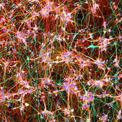
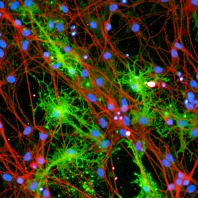
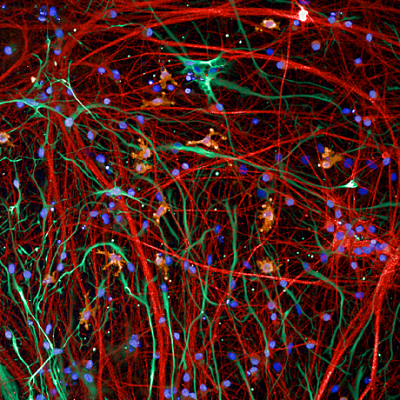
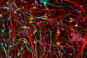
Representative immunofluorescent images showing (A) neurons (red, MAP2) and astrocytes (green, GFAP); (B) neurons (red, beta3 tubulin) and oligodendrocytes (MBP, green); and (C) neurons (red, MAP2), microglia (yellow, IBA1), and astrocytes (green, GFAP).